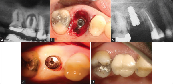
Figure 4.
(a) The maxillary right first molar suffered endodontic treatment failure and developed a large periapical lesion that had lifted the sinus floor via periosteal reaction. The inter-septal bone had been left intact coronally. (b) The inter-septal bone was sufficient to stabilize the implant but there remained both buccal and palatal bone dehiscence and large gaps. No grafting was done and the site allowed to heal by secondary intention. (c) A radiograph taken at 2 months showed new bone forming in the nongrafted socket. (d) This photo taken 3 months after implant placement showed excellent soft-tissue healing with minimal alveolar ridge remodeling. (e) The clinical situation 1 year after implant restoration showing excellent buccal tissue contours.

