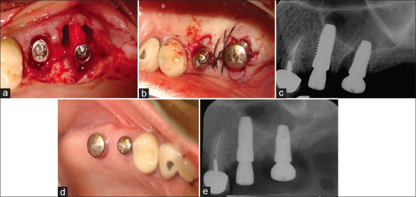
Figure 5.
(a) After atraumatic extractions, the clinician opted to place the molar implant in the palatal root socket leaving the two buccal roots un-grafted. (b) Minor flap manipulation was done to rest on and covers the undisturbed inter-septal bone. (c) The immediate postop radiograph shows the large defects left behind by not grafting the two buccal root sockets. (d) Excellent soft tissue healing was documented at 3 months. (e) A radiograph taken after 4 months healing.

