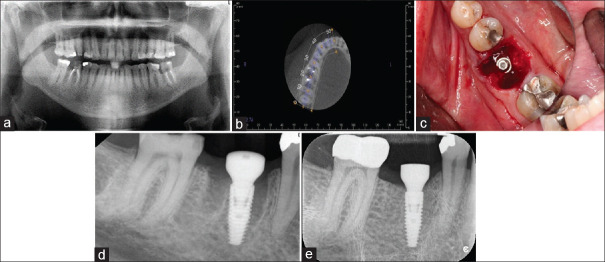
Figure 6.
(a) This mandibular right first molar was condemned as nonrestorable. (b) The axial slices of the cone-beam computed tomography scan suggested there to be adequate inter-septal bone to stabilize an IMI. (c) Once the implant had been placed, large gaps remained both mesially and distally. Surgery performed by Dr. Quang Nguyen, Periodontics Resident, Faculty of Dentistry, University of Toronto. (d) The immediate postoperatve radiograph confirms good implant positioning and use of a large diameter healing abutment. (e) A radiograph taken at 4 months revealing excellent bone healing.

