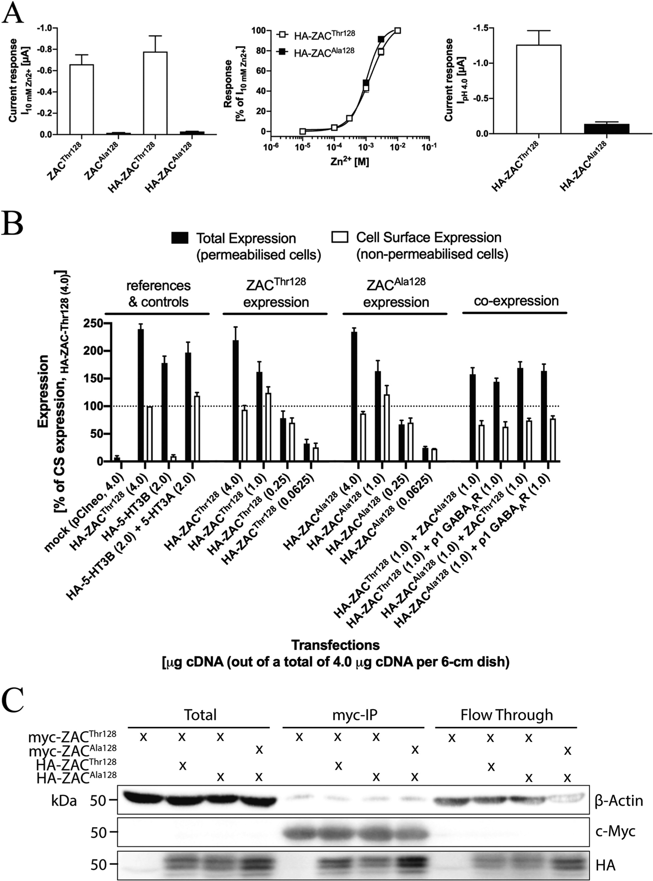
Fig. 8. Expression properties of ZACThr128 and ZACAla128 in tsA201 and HEK293 cells.

A. Functional properties of HA-ZACThr128 and HA-ZACAla128 expressed in oocytes injected with 1.84 ng cRNA of both subunits. Left: Averaged current amplitudes evoked by Zn2+ (10 mM) in ZACThr128-, ZACAla128-, HA-ZACThr128- and HA-ZACAla128-expressing oocytes (mean ± S.E.M., n = 4–8). Middle: Averaged concentration-response relationships displayed by Zn2+ at HA-ZACThr128 and HA-ZACAla128 (means ± S.E.M., n = 6–8). Right: Averaged current amplitudes evoked by H+ (pH 4.0) in HA-ZACThr128- and HA-ZACAla128-expressing oocytes (means ± S.E.M., n = 6–8). B. Total and cell surface expression levels of HA-ZACThr128 and HA-ZACAla128 transiently expressed in tsA201 cells in the ELISA experiments. All transfections used a total of 4 μg cDNA (all in the pCIneo vector) per 6-cm dish, where the quantities of specific cDNAs indicated in the figure were supplemented with “empty” pCIneo up to 4 μg. Total and cell surface expression was determined in permeabilized (Triton-X-treated) and non-permeabilized cells, respectively. Data (in %, normalised to the absorbance for the reference “HA-ZACThr128 (4.0)”-transfected cells) are given as mean ± S.E.M. and are based on a total of three experiments (n = 3). A line marker indicates the 100% level. C. Co-immunoprecipitation of ZACThr128 and ZACAla128. HEK293 cells were transfected with myc-ZACThr128 alone, myc-ZACThr128/HA-ZACThr128, myc-ZACThr128/HA-ZACAla128, or myc-ZACAla128/HA-ZACAla128. ZAC proteins were immunoprecipitated using myc-beads. Lysates and immunoprecipitated samples were resolved by SDS-PAGE. The myc-, HA- and actin-expression levels were measured by immunoblot.
