Figure 4.

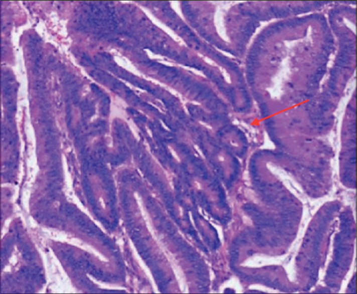
Representative H and E staining of malignant mucinous cystic lesions. Histology of cyst wall with papillary projections (red arrow)

Representative H and E staining of malignant mucinous cystic lesions. Histology of cyst wall with papillary projections (red arrow)