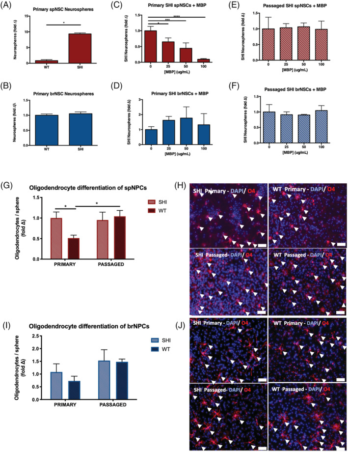
FIGURE 1.

Myelin basic protein (MBP) inhibits neural stem cell proliferation and oligodendrocyte differentiation through its interaction with the spinal cord niche. A, A 10‐fold increase in the number of spinal cord‐derived neurospheres grown from primary culture of SHI mice compared to wild‐type (WT) controls (n = 3 independent experiments; P = .0124). B, No difference in the number of forebrain‐derived neurospheres from SHI and WT control mice. C,D, The numbers of primary SHI neurospheres are reduced in the presence of exogenous MBP in a dose‐dependent manner in the spinal cord—25, 50, and 100 μg/mL—(C) but not the forebrain (D) (n = 3 independent experiments per region). E,F, Passaged SHI neurosphere‐derived cells from spinal cord (E) and forebrain (F) are unaffected by the presence of exogenous MBP—25, 50, and 100 μg/mL (n = 4 independent experiments per region). G, Primary spinal cord neurospheres from WT mice give rise to significantly fewer oligodendrocytes than spinal cord neurospheres from SHI mice (P = .0475). Following passaging, oligodendrogenesis is no longer inhibited from WT‐derived neurospheres (P = .0473); n ≥ 7 neurospheres per group. H, Representative images of differentiated primary and passaged spinal cord‐derived neurospheres from SHI and WT mice; scale bar = 100 μm. Arrowheads indicate DAPI+/O4+ oligodendrocytes. I, Primary and passaged forebrain‐derived neurospheres from WT and SHI mice give rise to similar oligodendrocyte formation; n ≥ 6 neurospheres per group. J, Representative images of differentiated primary and passaged forebrain‐derived neurospheres from SHI and WT mice; scale bar = 100 μm. Arrowheads indicate DAPI+/O4+ oligodendrocytes. Data are represented as means ± SEM. Statistics: A,B, t tests; C‐F, one‐way ANOVAs; G,I, two‐way ANOVAs. *P < .05, ***P < .001, ****P < .0001
