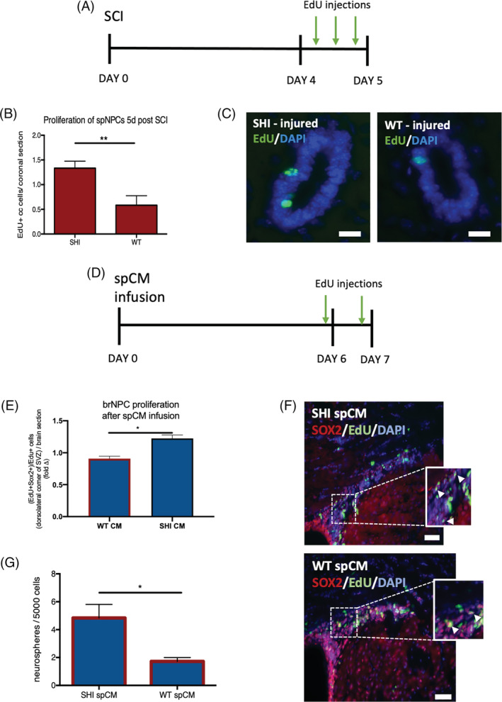
FIGURE 4.

Infusion of spinal cord‐derived conditioned media from wild‐type (WT) cultures inhibits neural precursor cell proliferation. A, Experimental paradigm; SCI, spinal cord injury, arrows = EdU injections. B, The numbers of EdU+ cells in the periventricular region of the spinal cord in WT mice is significantly reduced compared to SHI mice (n = 3 mice per group). C, Representative image of EdU+ cells (green) in spinal cord periventricular region at 5 days post SCI; scale bar = 20 μm. D, Experimental paradigm for the spCM intraventricular infusion. E, The fold change in SHI CM vs WT CM are shown relative to the Control media infused brains. There is a significant decrease in Sox2+/EdU+ NPC proliferation in the presence of WT spCM in the dorsolateral corner of the LV (P = .0152). Red outline indicates the presence of CM; n = 3 mice per group. F Representative images of Sox2+/EdU+ cells from SHI‐derived and WT‐derived spCM infused brains; scale bar = 100 μm. G, Significantly fewer neurospheres are formed from forebrain of mice that received WT spCM infusion (n = 3 mice/condition). Red outline represents the presence of spCM. Data are represented as means ± SEM. Statistics: B,E, t tests; G, one‐way ANOVA. *P < .05, **P < .01
