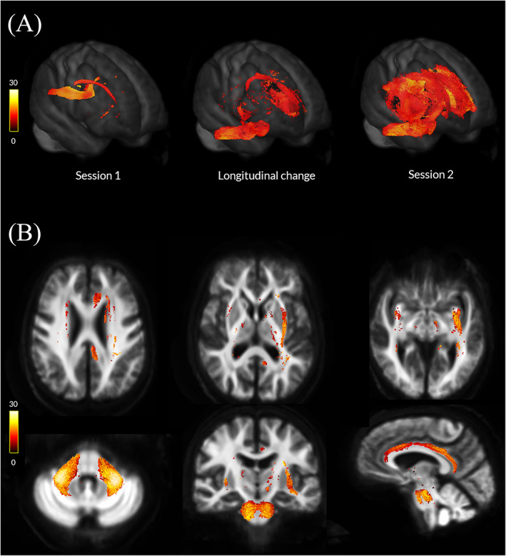
FIG. 2.

Macrostructural white matter changes in patients with Parkinson's disease and low visual performance over time. (A) Changes in white matter macrostructure (as seen by reduction in fiber cross‐section [FC]) in PD low visual performers compared with PD high visual performers (FWE‐corrected P < 0.05) in session 1 (baseline, left), at longitudinal change (difference between the 2 images, middle), and in session 2 (18 months follow‐up, right). Results are presented as streamlines and colored by percentage reduction. (B) Statistically significant (FWE‐corrected P < 0.05) longitudinal reductions in fiber cross‐section (FC) in PD low visual performers compared with PD high visual performers. Results are colored by percentage reduction. [Color figure can be viewed at wileyonlinelibrary.com]
