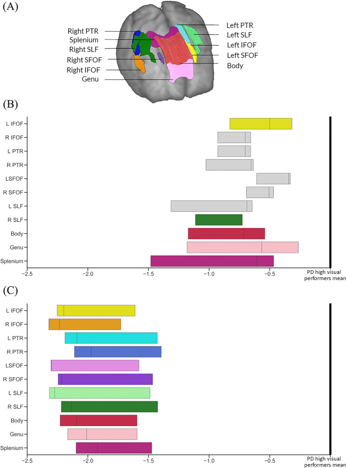
FIG. 4.

Significant tracts in Parkinson's low performers; tract of interest analysis. (A) Anatomical representation of all analyzed tracts. PTR, posterior thalamic and optic radiations; SLF, superior longitudinal fasciculi; IFOF, Inferior fronto‐occipital fasciculi (segmentation includes the inferior longitudinal fasciculus); and SLF, superior fronto‐occipital fasciculi. (B) Baseline visit. Reduction (mean, 95% CI) in fiber cross‐section (FC) visualized as percentage reduction from the mean of patients with Parkinson's disease with high visual performance. Tracts with significantly reduced FC (FDR‐corrected P < 0.05) are shown in color, whereas tracts with no significant changes in FDC are plotted in gray. L, left; R, right; C, visit 2 (18‐month follow‐up). Reduction (mean, 95% CI) in fiber cross‐section (FC) visualized as percentage reduction from the mean of patients with Parkinson's disease with high visual performance at follow‐up. All 11 of the selected tracts showed significantly reduced FC (FDR‐corrected P < 0.05). L, left; R, right. [Color figure can be viewed at wileyonlinelibrary.com]
