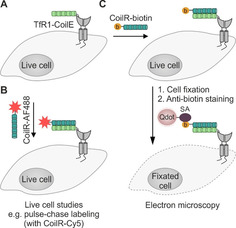
Figure 6.

Labeling of membrane proteins by using the VIPER system. A) The transferrin receptor 1 (TfR1, gray) is fused to the CoilE‐tag. B) Administration of the CoilR probe, equipped with an AF488 fluorophore (red star) enables visualization of the POI and live cell experiments. C) The CoilR can be equipped with a biotin group (yellow) to label the protein. Upon cell fixation, the biotin label is addressed by a streptavidin probe (SA, purple) quarrying a quantum dot (Qdot, light pink), allowing electron microscopy.
