Figure 10.

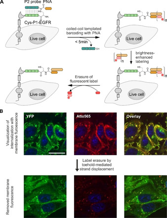
Labeling of membrane proteins by coiled‐coil‐templated barcoding with peptide nucleic acid (PNA). A) The POI (gray) was N‐terminally fused to the peptide tag Cys‐P1‐tag (light green). Addition of peptide probe P2 (jade green) triggers a coiled‐coil‐induced labeling reaction that installs a PNA tag. The PNA‐Cys‐P1‐POI is fluorescently labeled by hybridization with a complementary oligonucleotide (light brown bar) equipped with a fluorescent dye (red star). Brightness‐enhanced labeling of PNA‐Cys‐P1‐POI is achieved by hybridization with an elongated DNA strand, allowing multiple hybridization steps with smaller fluorescently labeled oligonucleotides (light red bar). Using toehold‐mediated strand displacement, the fluorescence is erased from the PNA by displacing the small fluorescent DNA fragments with complementary DNA sequences (dark red bar). B) EGFR was labeled with Atto565 by coiled‐coil‐induced PNA barcoding and DNA hybridization, and subsequently treated with EGF. It is difficult to detect internalized EGFR behind the background of EGFRs on the membrane (upper row). Internalized receptors are visualized with higher signal‐to‐noise ratios upon toehold‐mediated strand displacement (lower row). Microscopy data adapted from Gavins, et al. [89]
