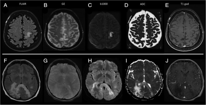
FIGURE 1.

Magnetic resonance imaging findings at the time of progressive multifocal leukoencephalopathy (PML) diagnosis in Patient 1, in A–E. and Patient 4, in F–L. (A) Patient 1 had a single progressive multifocal leukoencephalopathy (PML) lesion located in the left rolandic white matter, hyperintense on axial FLAIR images. (B) The adjacent cortex shows a characteristic rim of hypointensity on gradient echo (GE) images. (C, D) No diffusion restriction is evident on b1000 sequences, in C, and ADC sequences, in D, and (E) no contrast enhancement was evident on T1 sequences after gadolinium injection. (F) Patient 4 had a large PML lesion in the right parietal white matter that involved the corpus callosum splenium and reached the contralateral hemisphere, evident as hyperintense on FLAIR images. (G) No cortical rim of hypointensity was evident on GE images. (H, I) Linear rims of diffusion restriction were present along the lateral margins of the PML lesion, corresponding to the front of active demyelination. (J) The PML lesion showed marked hypointensity on T1 sequences after gadolinium injection, but no contrast enhancement. FLAIR, Fluid Attenuated Inversion Recovery.
