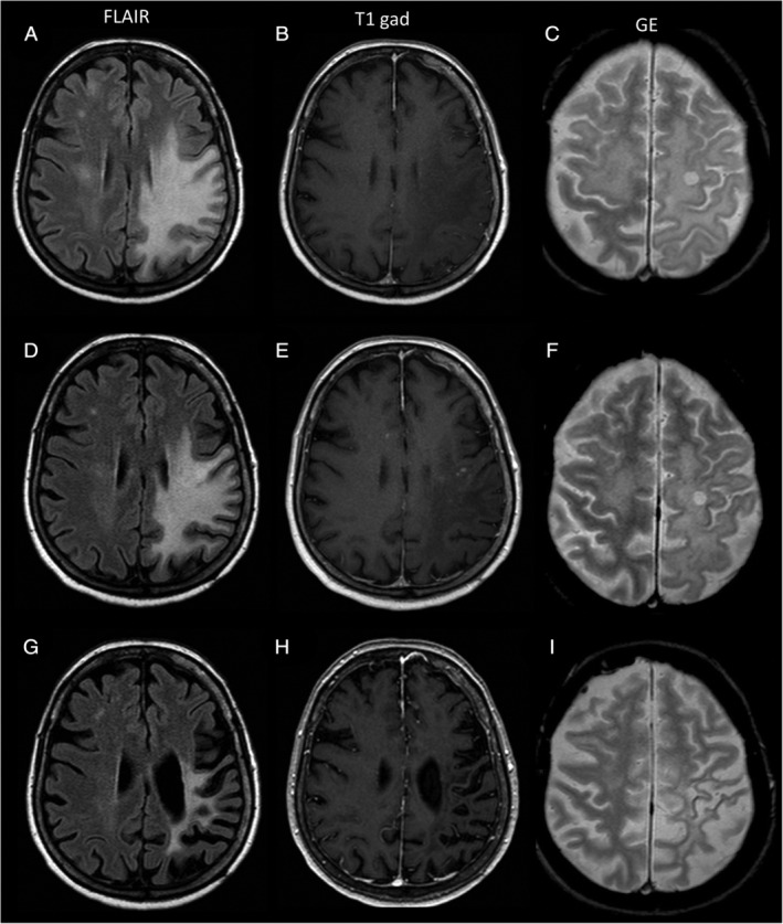
FIGURE 2.

Magnetic resonance imaging (MRI) findings in Patient 1. (A–C) Before starting T cell therapy. (D–F) 1 month after T cell therapy discontinuation. (G–I) 2.5 years after T cell therapy discontinuation. (A, D, G) Axial FLAIR images. (B, E, H) T1‐weighted images after gadolinium injection. (C, F, I) Gradient echo (GE) images. (A, B) MRI at initiation of T cell therapy showed a large frontoparietal progressive multifocal leukoencephalopathy (PML) lesion that was hyperintense on FLAIR sequences, shown in A, and hypointense on T1 sequences, with no contrast enhancement after gadolinium injection, shown in B. (D) MRI acquired 1 month after the last T cell therapy infusion showed an initial reduction in the size of the PML lesion on FLAIR images, together with a sharp reduction of its mass effect. (E) On T1 sequences after gadolinium injection, small punctate areas of contrast enhancement had appeared within (or close to) the PML lesion. (G, H) MRI acquired at the last follow‐up showed further reduction of the FLAIR hypersignal, in G, and a major cortical–subcortical atrophy across the left frontoparietal lobes, in G and H. FLAIR, Fluid Attenuated Inversion Recovery.
