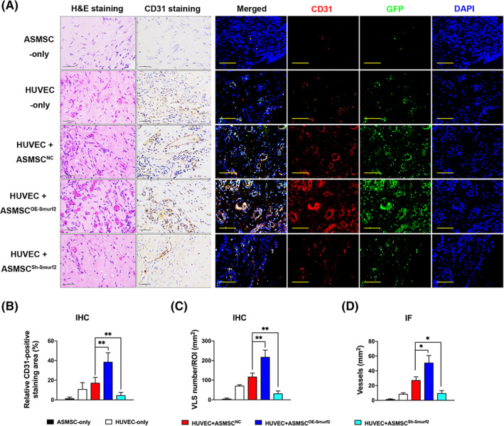
FIGURE 3.

Matrigel plug assay in vivo. We pretransfected human umbilical vein endothelial cells (HUVECs) with an empty lentivirus vector expressing the GFP protein to label the HUVECs and then marked these HUVECs with GFP staining to distinguish them from mouse endothelial cells and mesenchymal stem cells (MSCs). A, H&E and CD31 IHC staining (scale bar = 50 μm) and IF staining (scale bar = 100 μm; red, CD31; green, GFP; blue, DAPI) illustrating varying blood vessel morphologies and quantities in the implants from the five groups. Vessel‐like structures (VLS) were defined by the presence of CD31‐positive structures with lumens. Stained sections contained VLS and red blood cells in the implants from the HUVEC‐only group, HUVEC+ASMSCNC group, HUVEC+ASMSCOE‐Smurf2 group and HUVEC+ASMSCSh‐Smurf2 group. Compared with the HUVEC+ASMSCNC group, the HUVEC+ASMSCOE‐Smurf2 group exhibited many more newly formed blood vessels with clear lumens and borders, whereas the HUVEC+ASMSCSh‐Smurf2 group exhibited significant decreases in these parameters. B‐D, Quantification of the angiogenic capacity is presented as the vessel density and percentage of the CD31‐stained area. n = 3 different ASMSC lines per group. All data are presented as the means ± SD. *P < .05, **P < .01
