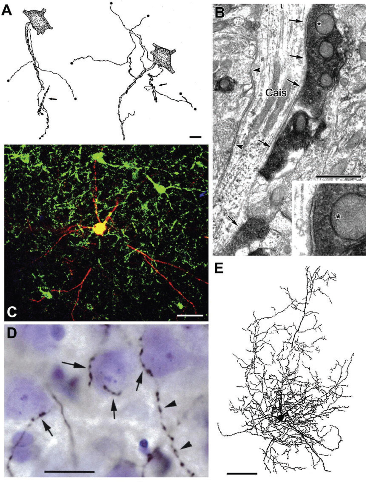
FIG. 5.

Axo-axonic cells (AACs) and basket cells in the rat BLa. (A) Drawings of two AAC axons whose axonal cartridges form multiple contacts with AISs of PNs (stippled). Asterisks indicate points at which collaterals continue but are not drawn. Each axon also forms additional axonal cartridges whose targets are not stained (arrows). (B) A CaMK+ axon initial segment of a PN (Cais), exhibiting particulate labeling, receives symmetrical synaptic inputs from densely stained PV+ axon terminals (arrows) and unstained PV− negative terminals (arrowheads). The upper PV+ terminal (asterisk) is shown magnified in the inset. (C) Merged confocal image of a biocytin-filled burst-firing PVBC. Biocytin is red. PV+ neurons are green. Yellow indicates double labeling (i.e., biocytin-filled structures that are PV+). (D) Photomontage of a Nissl-stained section showing a portion of the axonal arborization of the neuron shown in (C), after processing for the avidin-biotin peroxidase technique. Its biocytin-filled axonal collaterals (black) form multiple contacts with the somata of three presumptive PNs (arrows). The axon of this PV+ cell contacted the somata of over 100 surrounding Nissl-stained cells. The cell on the right receives additional axonal contacts along the proximal portion of a downwardly projecting process. This axon then continues its downward course (arrowheads) forming a series of varicosities that do not contact Nissl-stained structures. (E) Drawing of the burst-firing PVBC shown in (C) and (D). Only the dendrites and axonal branches in the 75-μm-thick section containing the cell body are drawn. Additional axonal and dendritic branches extended into two adjacent 75-μm-thick sections. Scale bars=10μm in (A), 1μm in (B), 50μm in (C), 20μm in (D), 100μm in (E). (A) Reproduced with permission from McDonald, A.J. (1982b). Neurons of the lateral and basolateral amygdaloid nuclei: A Golgi study in the rat. The Journal of Comparative Neurology, 212, 293–312. (B) Reproduced with permission from Muller JF, Mascagni F, McDonald AJ (2006) Pyramidal cells of the rat basolateral amygdala: Synaptology and innervation by parvalbumin-immunoreactive interneurons. The Journal of Comparative Neurology, 494, 635–650. (C)–(E) Reproduced with permission from Rainnie DG, Mania I, Mascagni F, McDonald AJ (2006) Physiological and morphological characterization of parvalbumin-containing interneurons of the rat basolateral amygdala. The Journal of Comparative Neurology, 498, 142–161.
