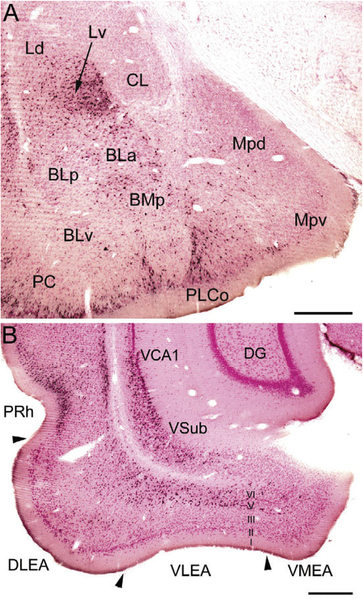
FIG. 8.

(A) Photomicrograph of retrogradely labeled neurons in the amygdala (black) at the bregma −3.1 level in a rat that received an injection of Fluorogold (FG) into the dorsolateral entorhinal area (immunoperoxidase technique with pink pyronin Y Nissl counterstain). The nomenclature used to denote the amygdalar nuclei is that used in the atlas by Paxinos and Watson (1997) (see Table 1). Additional abbreviations: Mpd, posterodorsal medial nucleus; Mpv, posteroventral medial nucleus; PC, piriform cortex. (B) Photomicrograph showing the locations of FG+ retrogradely-labeled neurons (black) in the ventral hippocampus and parahippocampal area at the bregma −6.3 level in a rat that received a large injection of FG into the BNC that involved both LA and BL (immunoperoxidase technique with pink pyronin Y Nissl counterstain). Note dense retrograde labeling in the perirhinal cortex (PRh), the deep layers (layers V and VI) of the dorsolateral and ventrolateral ERC (DLEA and VLEA, respectively), and the ventral subiculum (VSub) and adjacent ventral part of CA1 (VCA1), but not in the dentate gyrus (DG). Scale bars=500μm. (A) and (B) Reproduced with permission from McDonald, A.J., Zaric, V. (2015a). GABAergic somatostatin-immunoreactive neurons in the amygdala project to the entorhinal cortex. Neuroscience, 290, 227–242.
