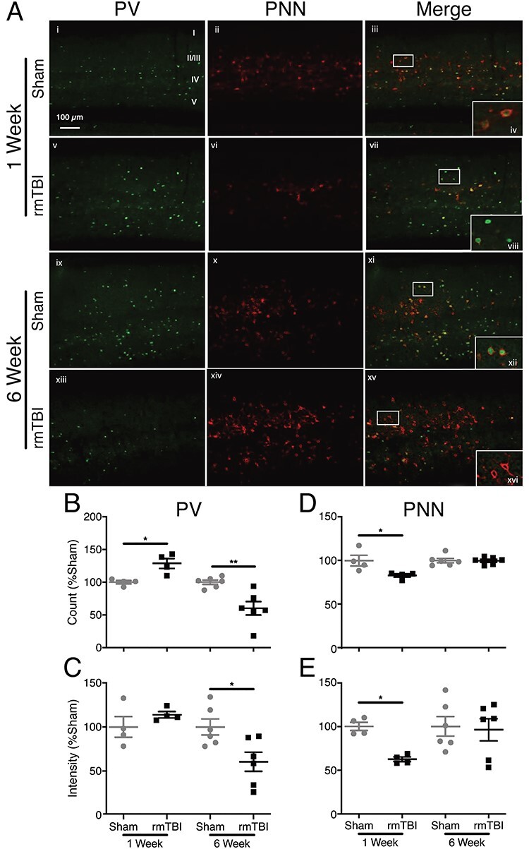
Figure 4 .

PV and PNN expression in barrel cortex. (A) IHC staining of barrel cortex. PV (green) and PNN (red). Callout top right healthy PVI enwrapped in healthy PNN. Callout second row PVI with degraded PNNs. Callout third row healthy PVI enwrapped in healthy PNN. Callout bottom “hollow” PNNs wrapping PVI that have lost PV expression. (B) There is an increase in PVI number 1 week after injury and a decrease in count 6 weeks after injury. (B) PV intensity does not change 1 week after injury; however, intensity decreases in injured mice at 6 weeks after rmTBI. (C) PNNs are reduced 1 week after injury and normalize by 6 weeks. (D) PNN intensity also decreases 1 week after rmTBI and normalizes 6 weeks after. (Bars indicate mean ± SEM, *P ≤ 0.05, **P ≤ 0.01).
