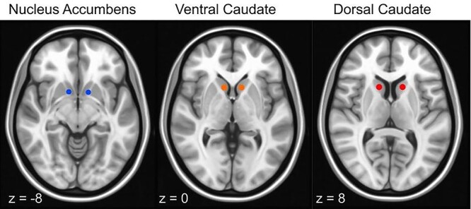
Figure 1 .

NAcc, ventral caudate, and dorsal caudate seed map: Image depicts nonoverlapping NAcc (blue) (right: x = 12, y = 10, z = −8; left: x = −10, y = 10, z = −8; only right is pictured); ventral caudate (orange) (bilateral, x = ±10, y = 15, z = 0); and dorsal caudate (red) (bilateral, x = ±13, y = 15, z = 9) seeds used to examine the strength of rsFC overlaid on MNI brain.
