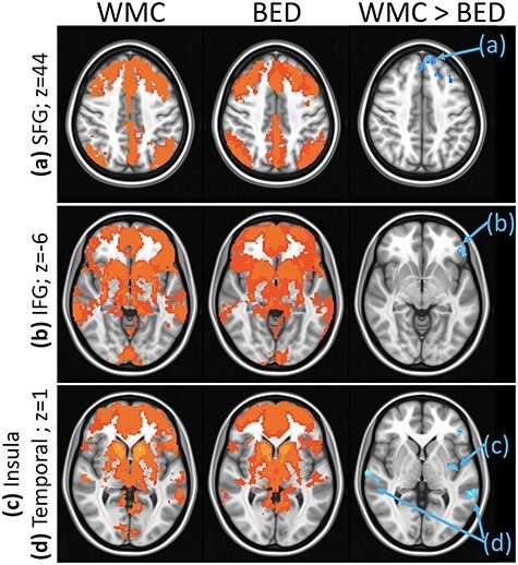
Figure 4 .

Group differences in rsFC from the dorsal caudate seed: Results showing higher rsFC between dorsal caudate and (a) left SFG (Brodmann area 8, axial slice z = 44), (b) left IFG (Brodmann area 47, axial slice z = −6), (c) left parietal lobule (Brodmann area 40, axial slice z = 1), (d) bilateral temporal gyrus (Brodmann areas 22 and 41, axial slice z = 1) in WMCs versus BED groups. Whole-brain rsFC maps showing regions with significant connectivity to dorsal caudate that survived thresholding and clustering to correct for multiple comparisons in WMC (first column) and BED (second column) groups. Third column shows whole-brain independent samples’ t-test results in which WMC had significantly higher rsFC than BED (p < 0.01, corrected for multiple comparisons). Functional maps are laid on MNI brains in radiological orientation, right (R) to left (L).
