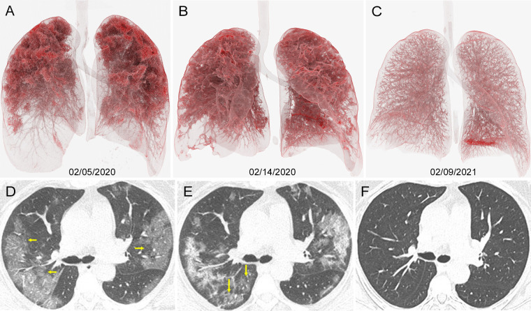
Images in 55-year-old woman diagnosed with severe COVID-19 with oxygen saturation of 73% at presentation. A–C, Cinematic renderings with, D–F, original CT scans help show evolution of her infection. A, D, Admission CT scans show confluent ground-glass opacities in both lungs (arrows in D), primarily distributed in upper lobes. B, E, CT scans 9 days later show progressive disease in lower lobes (arrows in E). C, F, Follow-up CT scans 1 year later show resolution of all previously seen lung findings.

An official website of the United States government
Here's how you know
Official websites use .gov
A
.gov website belongs to an official
government organization in the United States.
Secure .gov websites use HTTPS
A lock (
) or https:// means you've safely
connected to the .gov website. Share sensitive
information only on official, secure websites.
