. 2021 Jun 17;10:e68380. doi: 10.7554/eLife.68380

Figure 5. Charge distribution across the luminal entrance to the KDEL receptor binding pocket.

(a) KDEL receptor sequence alignment showing two regions centred around amino acid D50 and W120 of the human proteins. Cognate retrieval signal variants are shown to the right of the alignment. (b) The structure of the KDEL receptor with bound TAEHDEL highlighting key residues involved in ligand binding and variant residues D50, N54, and E117. (c) The charged surface for the WT KDEL receptor and (d) N50, N50/K54 and N50/K54/Q117 mutants is shown.

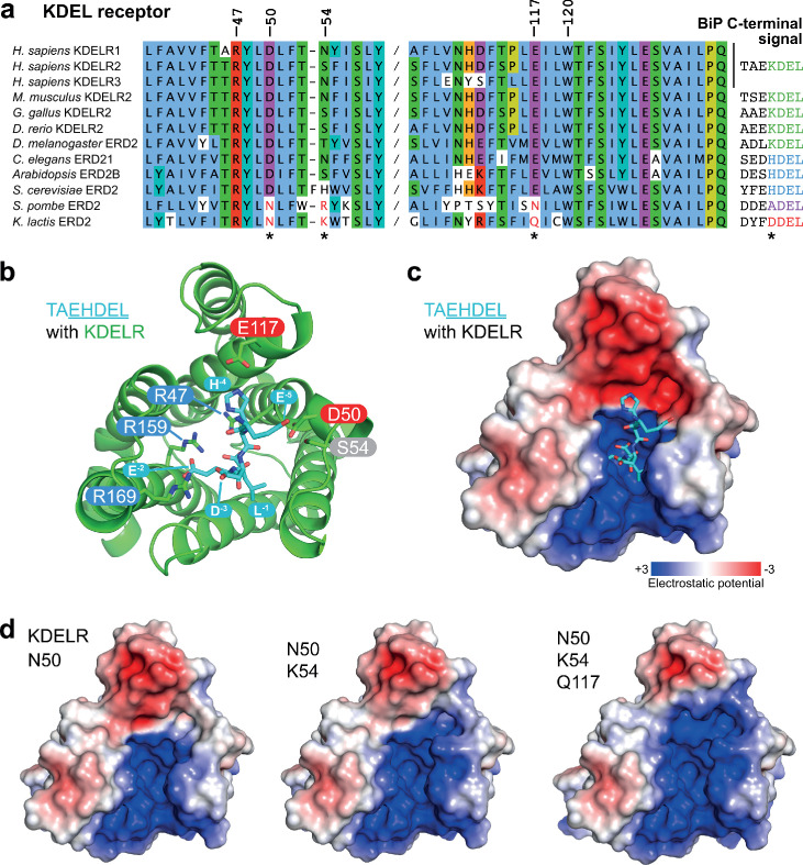
Figure 5.

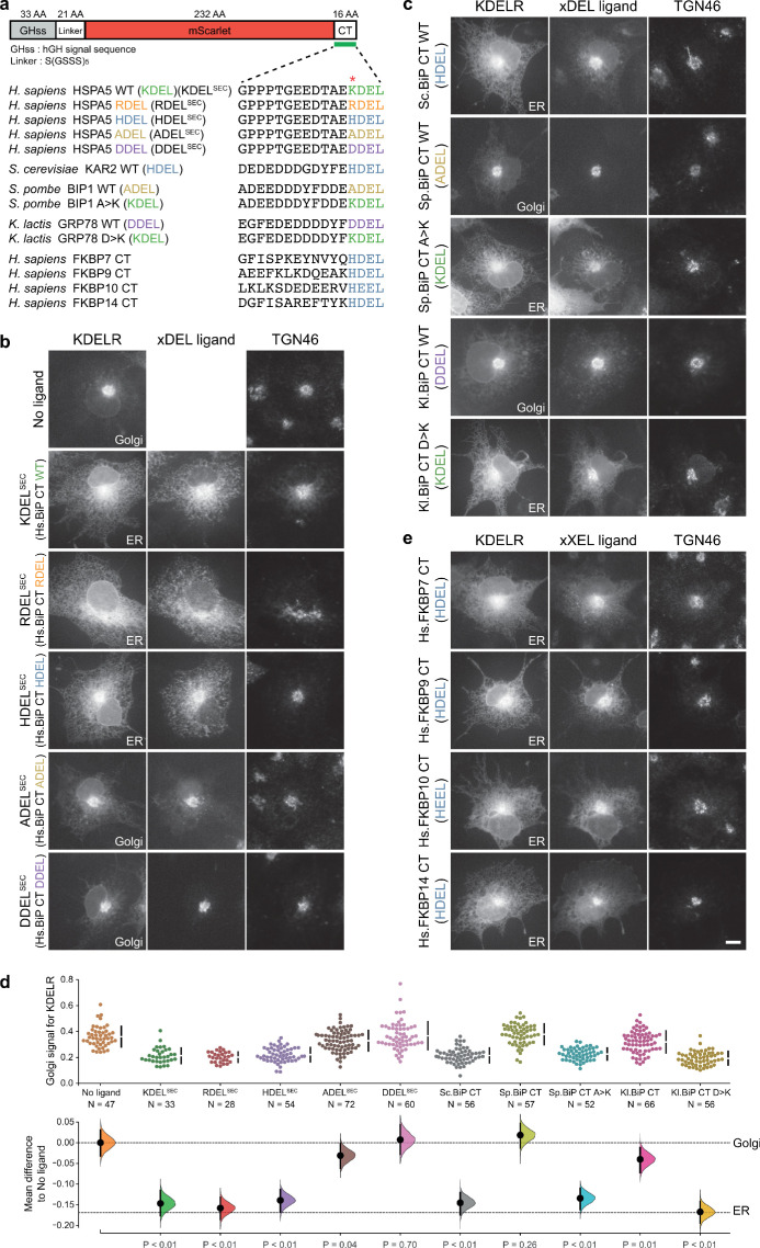
Figure 5—figure supplement 1. Comparison of human and yeast ER retrieval signals.

Figure 5—figure supplement 1.

(a) Schematic of the ER retrieval construct showing the human growth hormone signal sequence (hGHss), linker, mScarlet fluorescent protein, and 16 amino acid extension carrying a C-terminal (CT) retrieval signal from known human and yeast ER proteins. A sequence alignment shows the conservation of the retrieval signal. (b) WT KDEL receptor distribution was followed in the absence (-ligand) or presence of human BIP derived signals (K/R/H/A/DDELsec). (c) WT KDEL receptor distribution was followed in COS-7 cells in the absence (-ligand) or presence of yeast BIP derived signals K/R/HA/DDELsec. (d) The fraction of KDEL receptor localised to the Golgi was measured before (no ligand) and after challenge with the different retrieval signals tested in b. and c. Effect sizes are shown as the mean difference for retrieval signal comparisons against the shared -ligand control with sample sizes and p-values. (e) WT KDEL receptor distribution was followed in COS-7 cells in the absence (-ligand) or presence of ER retrieval signals from human FKBP family proteins. In all image panels, TGN46 was used as a Golgi marker and the scale bar is 10 µm.
Figure 5—figure supplement 1—source data 1. Source data for the ligand-induced KDELR receptor retrieval assays in Figure 5—figure supplement 1.