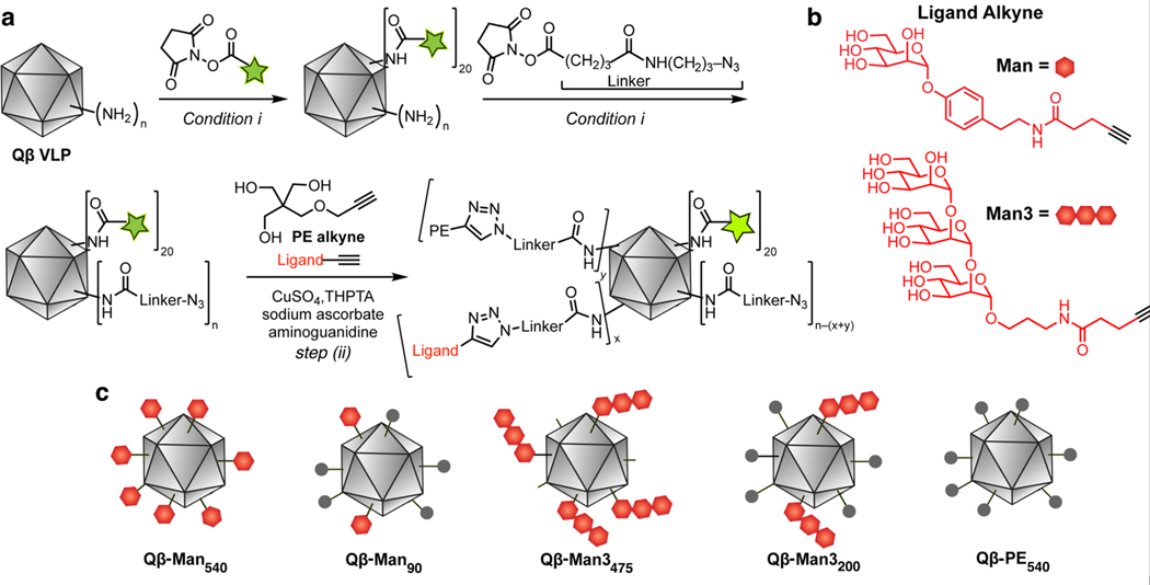
Figure 1. Preparation of fluorescently tagged Qβ particles.
Particles were generated that display an aryl mannoside (Man) group (red hexagon), a trimannoside group (3 linked hexagons), pentaerythritol monoether (PE, grey circle), or a combination. (a) Synthetic scheme (AF488 = Alexa Fluor 488). Conditions: (i) 0.1 M potassium phosphate buffer, pH 7.0, 0 °C to room temperature, 3 h. (step ii) tris-(3 hydroxypropyltriazolylmethyl)amine (THPTA), CuSO4, sodium ascorbate., aminoguanidine, 0.1 M potassium phosphate, pH 7.0, 37 °C, 4 h. (b) Structure of the attached DC-SIGN targeting groups. (c) Graphical representation of the functionalized Qβ VLPs.

