Figure 1.
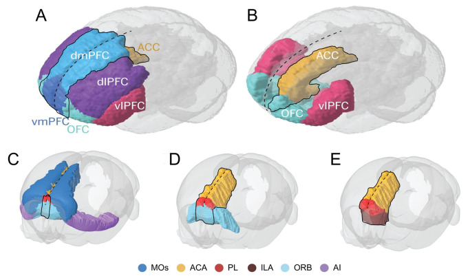
Functional divisions of the human, non-human primate and rodent (mouse) prefrontal cortex (A and B) Frontal-side view of the human primate brain with illustration of the prefrontal cortex functional divisions including the ACC, demarcated around the typically reported mPFC subregions of dmPFC, vmPFC and medial OFC. (C–E) Tilted frontal-side view of the rodent mouse brain illustrated with the agranular prefrontal cortex divisions and demarcated around the commonly stated mPFC subregions of ACA, PL, ILA and medial ORB. Dashed black line marks the sagittal midline. ACA, anterior cingulate area; ACC, anterior cingulate cortex; AI, agranular insular area; dlPFC, dorsolateral prefrontal cortex; dmPFC, dorsomedial prefrontal cortex; ILA, infralimbic area; MOs, secondary motor area; OFC, orbitofrontal cortex; ORB, orbital area; PL, prelimbic area; vlPFC, ventrolateral PFC; vmPFC, ventromedial prefrontal cortex. The schematic is adapted from Carlén.6

