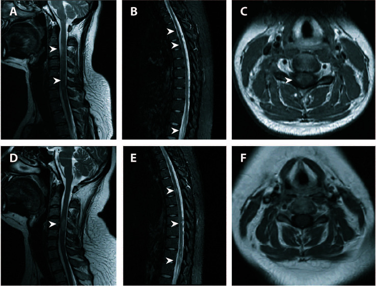
Figure 2.
Spinal MRI of the first and third attack. (A–C) Cervical (T2-weighted imaging) and thoracic (fat-saturated T2-weighted imaging) spinal MRI of the initial attack revealed multiple T2-hyperintense lesions throughout the spinal cord. Axial fat-saturated T1-weighted imaging with contrast enhancement showed eccentric lesions with patchy enhancement. (D–F) Repeat spinal MRI showed new lesions along the cervical and thoracic cord with resolution or attenuation of previous lesions. No enhancement was seen on axial fat-saturated T1-weighted imaging. Lesions were indicated by white arrowheads. MRI, magnetic resonance imaging.

