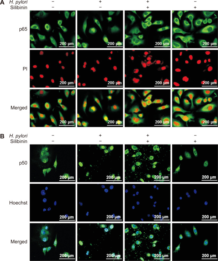
Figure 3. The effects of silibinin nuclear localization of p65 and p50 subuits of NF-κB.
Immunostaining visualizes the location of p65 (A) or p50 (B) protein in the nucleus stained with propidium iodide (PI) or Hoechst. MKN-1 cells were treated with Helicobacter pylori in the presence or absence of silibinin as described in Materials and Methods. Cells were fixed, permeabilized and stained with antibodies against p65 (A) and p50 (B) followed by treatment with 488-conjugated donkey anti-rabbit IgG. Nuclei were visualized by PI (A) or Hoechst (B). Magnification: ×400.

