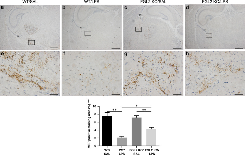
Fig. 1. Expression of MBP in WT and FGL2 KO mice brains after intrauterine inflammation.
MBP immunohistochemical staining was used to detect the myelination of white matter among the four groups on P7 (a–d low magnification, scale bars: 500 µm; e–h: high magnification, scale bars: 50 µm) (n = 6 for each group). On the hippocampus level, MBP positive staining was mainly observed in the periventricular region. The percentage of MBP positive staining area (e) are presented as mean ± SEM; *P < 0.05 and **P < 0.01.

