Fig. 3.
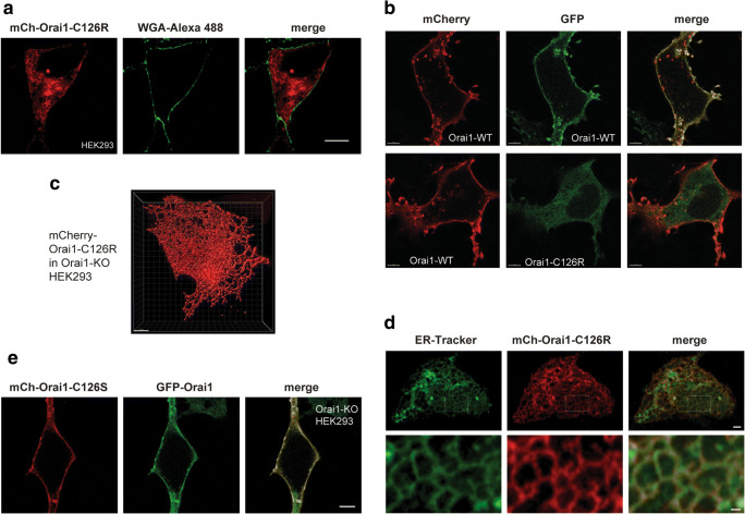
C126R mutation impairs ORAI1 PM localization. a Representative confocal airyscan of HEK293 cells expressing mCherry-ORAI1-C126R and co-stained with the PM marker WGA-Alexa 488. Scale bar 10 μm. b Confocal images of ORAI1-KO HEK293 cells co-expressing mCherry-ORAI1 (WT) with either GFP-ORAI1 (WT) or GFP-Orai1-C126R as indicated. Scale bar 5 μm. c 3D rendering of Orai1-C126R ER localization from a confocal z-stack of images from an Orai1-KO HEK293 cell transiently expressing mCherry-Orai1-C126R using the Imaris software. Scale bar 5 μm. d ORAI1-KO HEK293 cells transiently expressing mCh-ORAI1-C126R and stained with ER-tracker (green). Top panel scale bar 2 μm. The area in the white rectangle is magnified in the lower panels to highlight the reticular ER pattern. Scale bar 0.5 μm. e Representative confocal images of a cell co-expressing mCherry-ORAI1-C126S with GFP-ORAI1 (WT). Scale bar 5 μm

