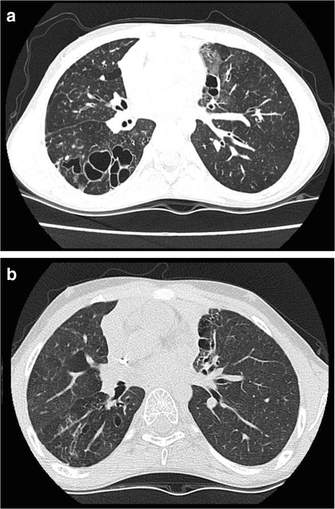
Fig. 3.

Thoracic computerized tomography images from patient 4. a Images taken 1 year pre-transplant showing markedly abnormal lung parenchyma with large cysts, bullae, and cystic bronchiectasis particularly in the right lower lobe in association with cylindrical bronchiectasis, bronchial wall thickening, bronchocoeles, and reduced lung attenuation. b Images taken 1 year post-transplant showing several thin walled pulmonary “cysts,” similar to previous pre-transplant findings and are therefore most likely to result from previous infection and lung destruction. Some regions of parenchymal distortion and scarring are also similar. There is some bronchial dilatation and distortion in regions of scarring, but no convincing evidence of bronchiectasis
