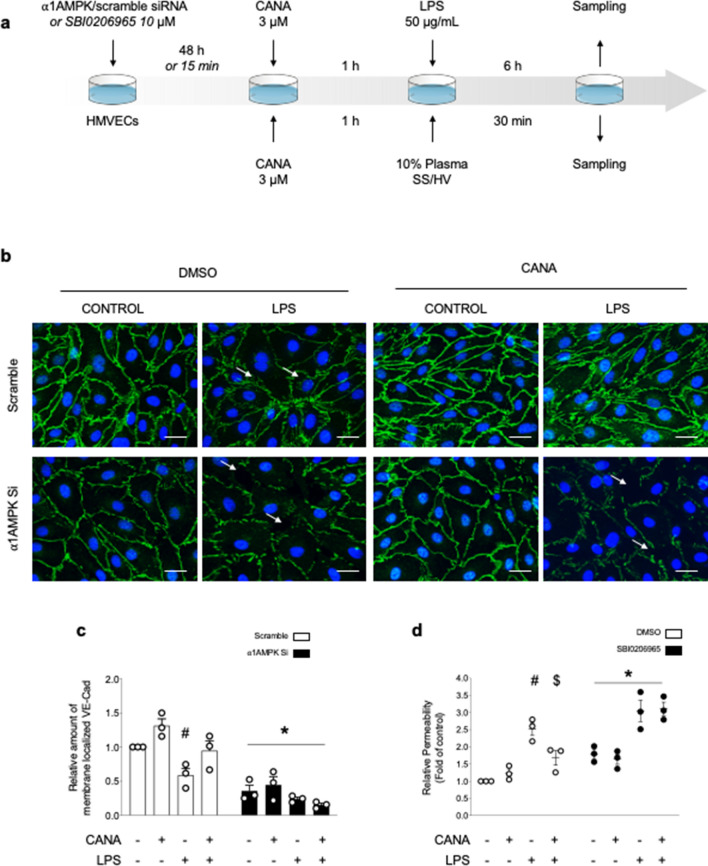
Figure 4.
AMPK activation by Canagliflozin protects VE-Cad organization and endothelial barrier function against LPS injury. (a) Schematic representation of the experimental protocol. For (b, c), HMECs were transfected with scramble or ⍺1AMPK targeting siRNA (50 nM) for 48 h, then treated with canagliflozin (Cana) (3 µM) or DMSO for one hour, before adding lipopolysaccharide (LPS) or vehicle (50 µg/mL) for 6 h. For (d), SBI compound was incubated for 15 min, then canagliflozin (3 µM) or DMSO were incubated for one hour, before adding LPS (50 µg/mL) for 6 h. (b, c) VE-Cad immunostainings were performed on HMECs treated according to the protocol detailed in (a). Representative images (b) and quantifications (c) are shown. Intercellular gaps are indicated by white arrows. Nuclei were stained with DAPI. Scale bar, 50 µm. (d) Endothelial permeability in response to canagliflozin, LPS challenge, and AMPK inhibitor SBI0206965. HMECs were grown on gelatin-coated Transwell inserts for 72 h and treated according to the protocol detailed in (a). Data are expressed as mean ± SEM (3 biological replicates for each condition). #p < 0.05 is relative to respective non-treated HMECs, $p < 0.05 is relative to LPS-only treated HMECs, and *p < 0.05 is relative to cells treated with DMSO. The data underwent two-way ANOVA.

