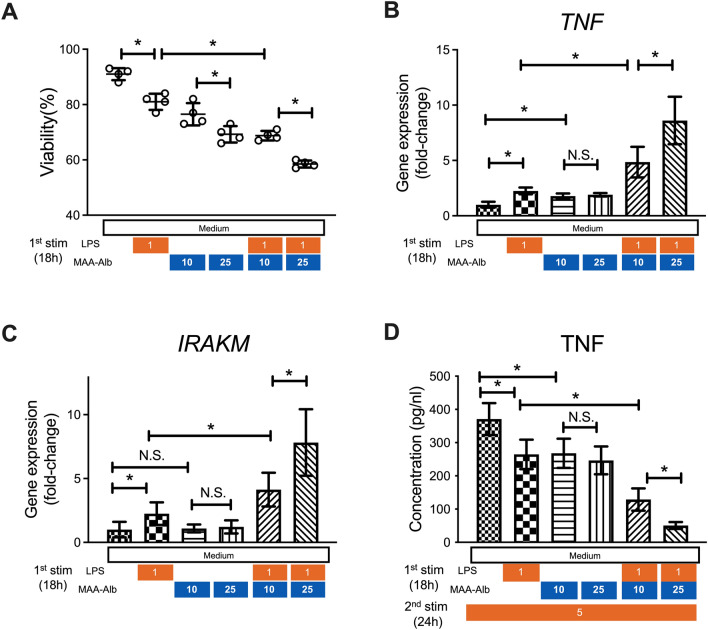
Figure 4.
MAA-Alb enhances LPS stimulation of CD14+ monocytes, and cells exposed to MAA-Alb produce less TNF-α upon secondary LPS stimulation. CD14+ monocytes collected from healthy donors were stimulated in the presence of LPS (1 ng/mL) and Alb (10–25 µg/mL) or MAA-Alb (10–25 µg/mL) for 18 h in vitro. (A–C) Viability of (A) and gene expression of TNF-α (B) and IRAK-M (C) in monocytes following stimulation. Cells were stimulated with 5 ng/mL LPS for 24 h. (D) Concentration of TNF-α in the culture supernatants of the monocytes following the second LPS stimulation for an additional 24 h in vitro. Data represent the mean ± SD. *p < 0.05, as determined by one-way ANOVA, N.S.: not significant.

