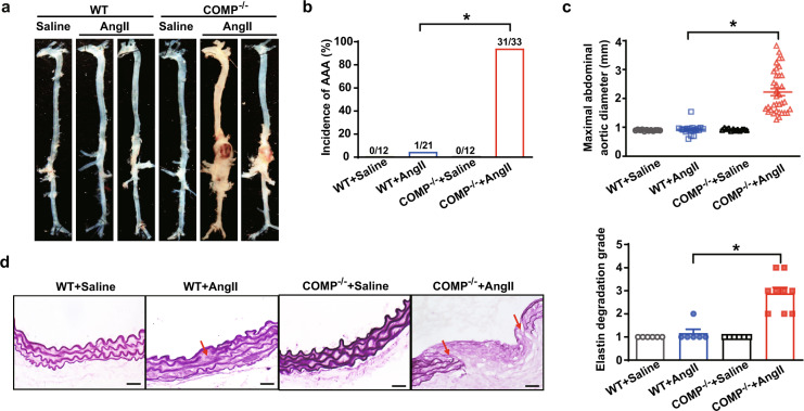
Fig. 2. COMP deficiency aggravates AngII-induced AAA formation.
a Representative images of the morphology of whole aortas from WT C57BL/6 J and COMP–/– mice with or without 28 days of AngII infusion. b Incidence of AngII-induced AAA in WT (n = 21) and COMP–/– mice (n = 33). *P < 0.05 by χ2 test. c The maximal abdominal aortic diameters (WT + Saline, n = 12; WT + AngII, n = 21; COMP–/– + Saline, n = 12; COMP–/– + AngII, n = 33). *P < 0.05 by Kruskal-Wallis test followed by Dunn’s test. d Representative images of Gomori staining and quantification of elastin degradation (WT + Saline, n = 6; WT + AngII, n = 6; COMP–/– + Saline, n = 6; COMP–/– + AngII, n = 9). *P < 0.05 by Kruskal-Wallis test followed by Dunn’s test. Scale bars, 50 μm.

