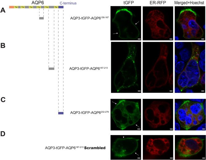
Figure 3.
AGR scanning of the second half of AQP6. (A) Approaching the second half of AQP6 in a similar manner to Fig. 2, peptide residues AQP6158–167 when C-terminally attached to AGR results in a construct that reaches the PM (white arrows). (B) However, when residues AQP6187–213 are C-terminally attached to AGR, the construct does not reach the PM. (C) Although, AQP6232–276 residues (compromising AQP6 C-terminal) attached to AGR does localize to the PM. (D) Interestingly, when a scrambled version of peptide residues AQP6187–213 are substituted for the native AQP187–213 and C-terminally attached to AGR, this construct is able to reach the PM. Cells were observed microscopically using an upright Olympus BX51WI equipped with 100 × 1 NA objective and images were captured using a Grasshopper3 CMOS camera (FLIR, Richmond, BC, Canada) controlled by Leica LAS X version 3.5 software (available at: https://www.leica-microsystems.com/products/microscope-software/p/leica-las-x-ls/). All figures were created using Adobe Illustrator CS6 (Available at: https://adobe.com/products/illustrator), under an Adobe Inc., Creative Cloud Desktop 2019 shared device license to Case Western Reserve University (CWRU) that operates until 3/31/2022. Scale bars: 8 µm.

