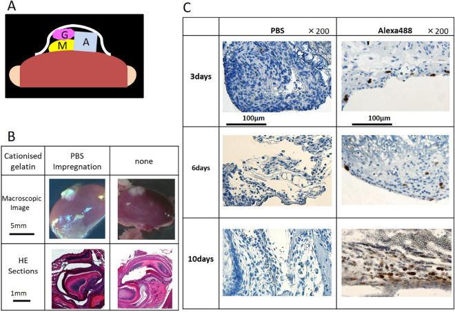
Figure 2.
Renal capsule assays using lateral mandibles from wild-type mice. (A) Schematic illustration of the procedure used for renal capsule transplantation. M indicates the lateral mandibles of E10 mice, G indicates cationized gelatin containing Stealth siRNA, and A indicates agarose agar (was used to maintain the graft space). (B) We evaluated H&E-stained sections, where the lateral mandible from wild-type mice was transplanted beneath the kidney capsule of nude mice (KSN/Slc). Renal capsule assays were performed with two groups: lateral mandibles transplanted with or without a cationized gelatin sheet containing PBS. Based on histochemical staining, we observed that the sections contained tooth structures, although we found no differences in the results with or without cationized gelatin. (C) Histological sections at days 3, 6, and 10 post -transplantation. Sections were evaluated by immunostaining. Magnification, 200× . Cells were immunostained with an Alexa Fluor 488-conjugated antibody.

