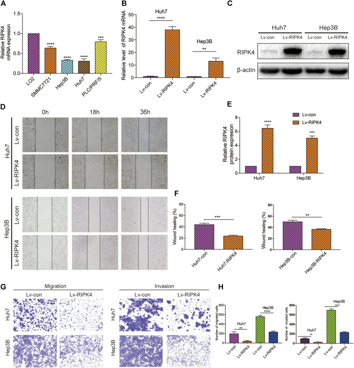
FIGURE 3.
RIPK4 inhibits the migration and invasion of HCC cells. (A) Real-time quantitative PCR analysis of RIPK4 mRNA levels in HCC cells compared to L02 cells. (B) Real-time quantitative fluorescent PCR analysis of RIPK4 expression after lentivirus transfection. (C) Western blotting analysis of RIPK4 expression after lentivirus transfection, and ß-actin was used as a control. (D) The wound healing showed that the migrated ability of Huh7-RIPK4 and Hep3B-RIPK4 cells was decreased. (E) Statistical analysis of three independent data of western blotting experiments. (F) Statistical analysis of three independent data of wound healing experiments. (G) Representative images (100 times) of Huh7 and Hep3B cell line Transwell migration and Matrigel invasion experiments. (H) Statistical analysis on three independent data of Transwell experiment. All experiments were performed three times independently, and the data are expressed as mean ± SD. **p < 0.01; ***p < 0.001.

