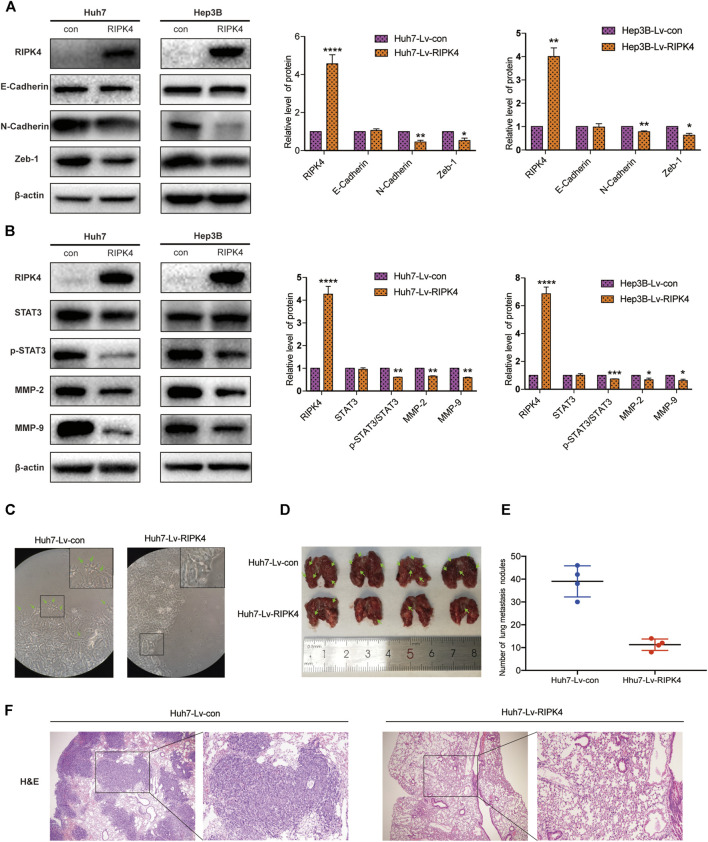
FIGURE 4.
RIPK4 inhibits EMT progression and STAT3 pathway activation in HCC. (A) Western blotting analysis of the expression levels of E-Cadherin, N-Cadherin, Vimentin, Zeb-1 protein in Huh7 and Hep3B cells infected with Lv-con and Lv-RIPK4 lentivirus, and ß-actin was used as a control. (B) Western blotting analysis of STAT3, p-STAT3, MMP-2, MMP-9 protein expression levels in Huh7 and Hep3B cells infected with Lv-con and Lv-RIPK4 lentivirus, and ß-actin was used as a control. (C) Representative images of huh7 morphology with Lv-con and Lv-RIPK4 lentivirus. (D,E) Representative images of lung metastases in mice and statistical analysis of the number of metastases. (F) Representative H&E staining pictures of mouse lung metastatic nodules. The data are expressed as mean ± SD. **p < 0.01; ***p < 0.001.

