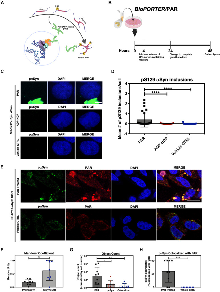
FIGURE 1.
Poly (ADP-ribose) (PAR) colocalizes with pαSyn. (A) Proposed mechanism of PAR-induced pαSyn accumulation, whereby PARP-1 hyperactivation due to DNA damage results in excess PAR production, leading to cytoplasmic pαSyn accumulation and pathogenic PAR–pαSyn interactions. (B) Experimental scheme of BioPORTER-mediated transfection of PAR polymer into SH-SY5Y-αSyn neuroblastoma cells. (C) Representative immunostain of pαSyn (green) and DAPI (blue) in SH-SY5Y-αSyn cells 48 h post treatment with either 50 nM PAR or ADP-HDP vs. BioPORTER alone (vehicle control). Scale bar 5 μm. (D) Quantification of pαSyn inclusions (aggregates larger than 1 μm) in PAR treated, ADP-HDP treated, and BioPORTER alone (vehicle control) samples. Bars represent mean ± SD. Two-way ANOVA followed by Tukey’s post hoc test (n = 3). ****P < 0.0001. (E) Representative IF immunostain of pαSyn (green), PAR polymer (red), and DAPI (blue) in SH-SY5Y-αSyn cells 48 h post treatment with 50 nM PAR vs. BioPORTER alone (vehicle control). Scale bar 10 μm. (F) Manders’ overlap coefficient analysis between total PAR over pαSyn inclusions (PAR/pαSyn) and pαSyn inclusions over total PAR (pαSyn/PAR) in the PAR treated samples, whereby an overlap coefficient of 0.5 implies that 50% of both objects (i.e., pixels) overlap. Bars represent mean ± SD. Student’s two-tailed t-test (n = 3). **P < 0.002. (G) Average object count in the PAR treated samples for the following objects: PAR, pαSyn inclusions, and colocalized PAR-pαSyn inclusions. Object counts were normalized by DAPI signal (i.e., cell number). Bars represent mean ± SD. Two-way ANOVA followed by Tukey’s post hoc test (n = 3). *P < 0.02, **P < 0.0045. (H) Colocalization analysis comparing the number of pαSyn inclusions colocalized with PAR immunostain in PAR treated and BioPORTER alone (vehicle control) samples. Images were captured using Zeiss LSM 710 confocal (40×/1.4 Oil) microscope. Bars represent mean ± SD. Student’s two-tailed t-test (n = 3). ***P < 0.0005. Graphical symbols represent fields-of-view containing 50–70 cells each.

