FIGURE 1.

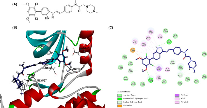
A, Chemical structure of YTH‐60. B, Docking of YTH‐60 into FGFR1 kinase X‐ray crystal structure in the three‐dimensional structure (PDB ID:5UR1). C, A two‐dimensional interaction map of YTH‐60 and FGFR1

A, Chemical structure of YTH‐60. B, Docking of YTH‐60 into FGFR1 kinase X‐ray crystal structure in the three‐dimensional structure (PDB ID:5UR1). C, A two‐dimensional interaction map of YTH‐60 and FGFR1