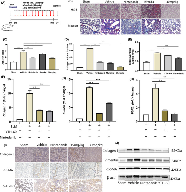
FIGURE 6.

YTH‐60 alleviates BLM‐induced pulmonary fibrosis in mice. A, The experimental schedule of YTH‐60 treatment for BLM‐induced pulmonary fibrosis. B, Representative images of YTH‐60 ameliorated the histopathological changes. Pulmonary tissues stained with H&E or Masson staining were shown from sham, BLM, BLM + YTH‐60 (15, 30 mg·kg−1) and BLM + Nintedanib (30 mg·kg−1) group. C, YTH‐60 treatment reduced the pulmonary fibrosis Ashcroft scoring. D, Morphometric analysis of collagen was used to measure the degree of lung fibrosis. E, Hydroxyproline content of lung tissues. Data are presented as mean ± SD, n = 3‐8 for each group, *P < .05; **P < .01; ***P < .001. F‐H, The mRNA levels of Collagen Ⅰ,α‐SMA and TGF‐β1 in the lung tissues was measured by Quantitative real‐time qPCR. Data are expressed as the mean ± SEM (n = 3 mice per group). I, Immunohistochemical determination of Collagen Ⅰ, α‐SMA and p‐FGFR expression in the lung tissues. J, The collagen Ⅰ, α‐SMA and vimentin of lung tissues were performed by western blotting
