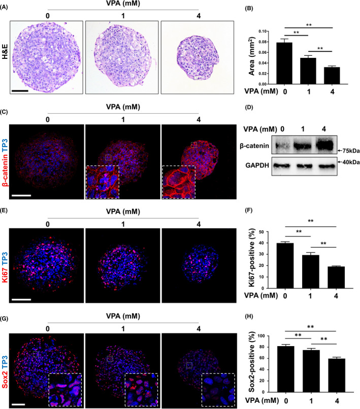
FIGURE 5.

Suspended culture of AM‐1 cells and effects of VPA on the growth. A‐H, Two‐hundred‐thousand AM‐1 cells were plated on a well of a low‐attachment surface 6‐well cell culture plate and cultured for 7 d in DMEM with or without VPA. Sections of spheroids were subjected to H&E staining (A), immunohistochemistry using antibodies to β‐catenin (C, red), Ki67 (E, red) and Sox2 (G, red); and immunoblotting using antibodies to β‐catenin or GAPDH (D). Nuclei were visualized using (C, E and G, blue). Scale bar = 200 μm. The size of sectioned spheroids (B) and ratio of Ki67‐positive (F) or Sox2‐positive (H) nuclei were quantified. n = 50. **p ‐value < 0.01. VPA, valproic acid, TP3, TO‐PRO‐3
