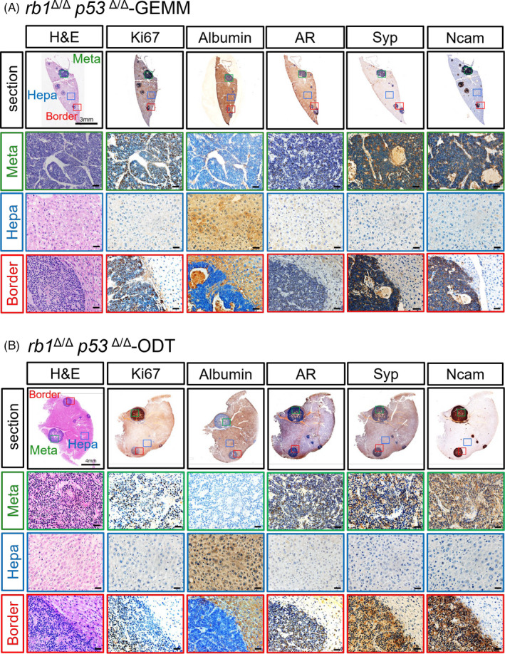
FIGURE 3.

Establishment of a rapid liver metastasis model for PCa in immune‐sufficient mice using rb1Δ/Δp53Δ/Δ GEMM organoids. A‐B, H&E and IHC staining images show the metastatic tumor foci in a liver lobe of rb1Δ/Δp53Δ/Δ GEMM mice (A) and rb1Δ/Δp53Δ/Δ GEMM organoids implanted mice (B). The metastatic region (meta, green), the border between the metastasis and the hepatocytes (border, blue) and the hepatic region (hepa, red) in the liver sections are shown. Ki67 antibody stains the proliferating cells of the tumor foci. Albumin is used as a marker of hepatocytes. AR antibody labels the tumor cells originated from the PCa. Syp and Ncam are used as markers for PCa cells with neuroendocrine differentiation phenotype
