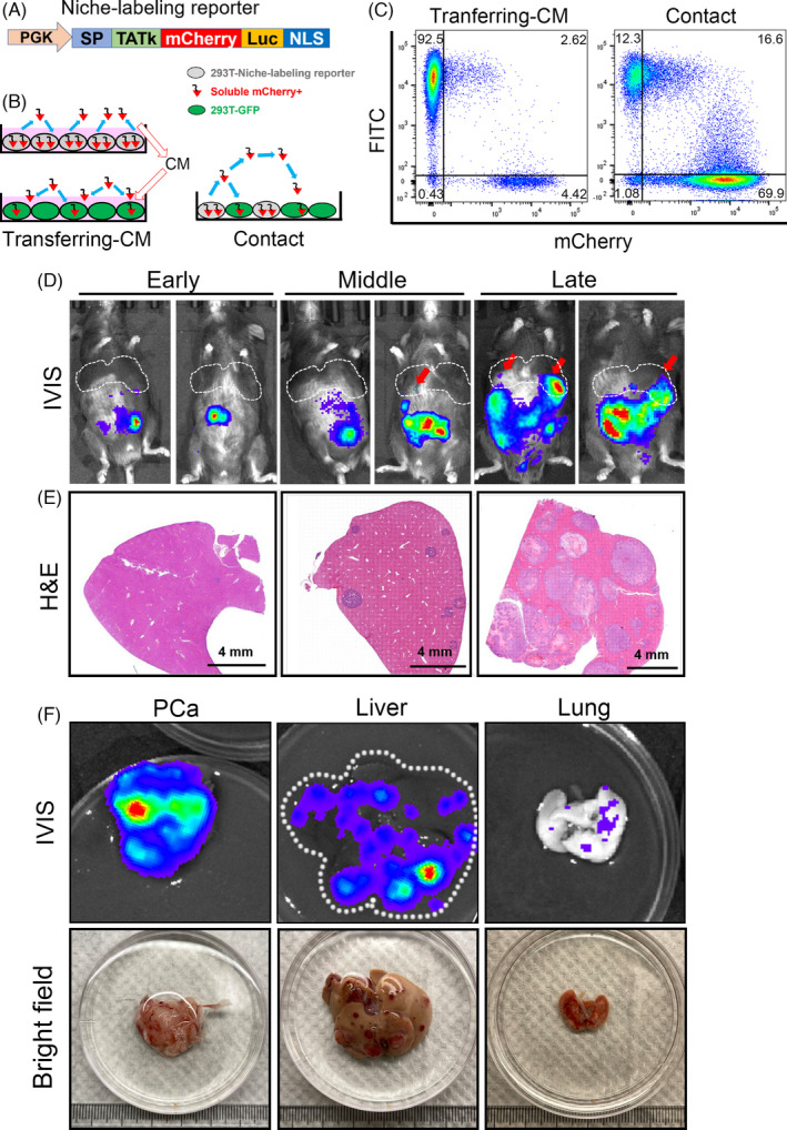
FIGURE 4.

A niche‐labeling system to reveal tumor‐niche communications in the liver metastasis of rb1Δ/Δp53Δ/Δ ODT mouse model. A, A lentiviral construction containing the PKG promoter driven transactivator of transcription (TATk) peptide followed by a secreted mCherry protein and a bioluminescent luciferase cassette. B, Proof of concept experiment of the niche labeling system using conditioned‐medium (CM) or directly contacted co‐culture method of 293T cells. C, Flow cytometry results show that the fluorescent mCherry protein can be secreted by mCherry+ 293T cells and uptaken by the GFP+ 293T cells. GFP+ 293T cells were cultured by transferring‐CM of mCherry+ 293T cells or directly co‐cultured with mCherry+ 293T cells in a contact manner. D, The bioluminescent images of rb1Δ/Δp53Δ/Δ ODT mice at early (20 d after inoculation), middle (35 d after inoculation) and late stages (50 d after inoculation). The liver metastatic tumor foci are indicated by red arrows. Rb1Δ/Δp53Δ/Δ GEMM organoids are infected with the niche‐labeling lentiviruses and orthotopically inoculated in the prostate of WT C57BL/6 mice (8‐weekold). E, H&E staining shows the liver metastatic foci in liver at early, middle, and late stages. Scale bar = 4 mm. F, The bioluminescent images and bright field images of PCa, liver and lung isolated from rb1Δ/Δp53Δ/Δ ODT mice
