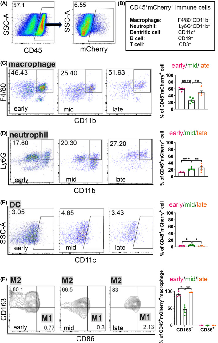
FIGURE 5.

Immune‐suppressive CD163+ macrophages are significantly increased in the metastatic niche at the late stage of PCa liver metastasis. A, Flow cytometric analysis of immune cells that are mCherry+ in the liver metastasis of rb1Δ/Δp53Δ/Δ ODT. B, A schematic of immune cells examined in the liver metastatic niche. C‐F, Representative flow cytometric plots and quantification of the ratio of macrophages (C), neutrophils (D) and dentritic cells (DCs) (E) in CD45+ mCherry+ cells, and M1, M2 macrophages (F) in CD45+ mCherry+ macrophages in rb1Δ/Δp53Δ/Δ ODT liver metastasis niche at the early, middle and late stages. Unpaired two‐tailed Student's t‐test is used for statistical analysis. The data are presented as the means ± SEMs. *P < .05, **P < .01, ***P < .001, ****P < .0001, n = 5
