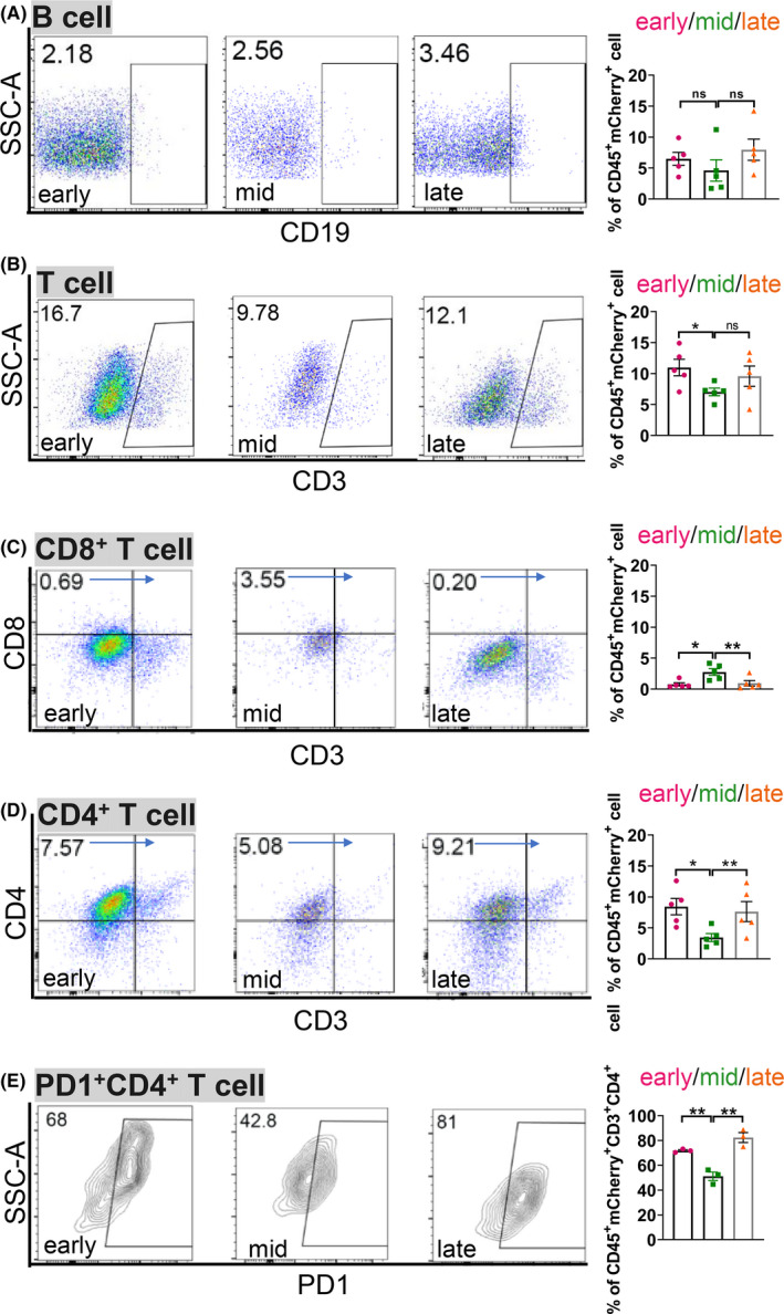
FIGURE 6.

A paucity of CD8+ T cells and abundant PD1+ CD4+ T cells further contribute to the immunesuppressive liver metastatic niche in rb1Δ/Δp53Δ/Δ ODT. A‐E, Representative flow cytometric analysis and quantification of the ratio of B cells (A), CD3+ T cells (B), CD8+ T cells (C) and CD4+ T cells (D) in CD45+ mCherry+ cells, and PD1+ CD4+ T cells (E) in CD45+ mCherry+ CD4+ T cells in rb1Δ/Δp53Δ/Δ ODT liver metastasis niche at the early, middle, and late stages. Unpaired two‐tailed Student's t‐test is used for statistical analysis. The data are presented as the means ± SEMs. *P < .05, **P < .01, n = 5
