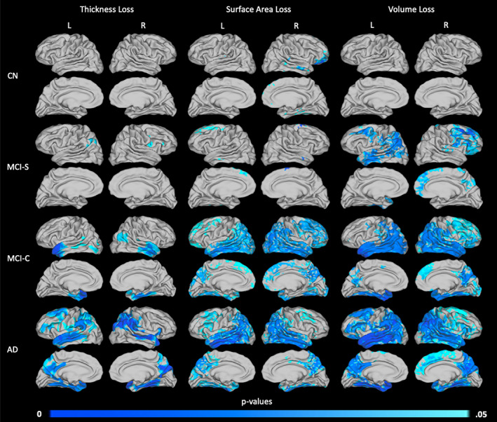
FIGURE 4.

Areas of significant thickness, surface area, and volume loss over a 2‐year period by diagnostic group. Patterns of atrophy are shown for the CN (n = 12, top row), MCI‐S (n = 12, second row), MCI‐C (n = 13, third row), and AD (n = 13, bottom row) groups, in terms of loss in thickness (left column), surface area (middle column), and volume (right column). p values are threshold‐free cluster‐enhanced with family‐wise error correction
