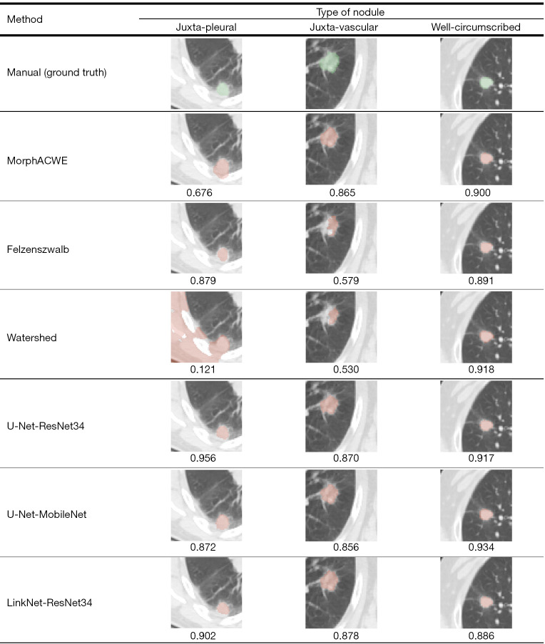
Figure 8.
Qualitative analysis of the segmentation results obtained with the three best-performing conventional and deep learning methods (Tables 3,4) on juxta-pleural, juxta-vascular and well-circumscribed nodules. The green overlays indicate manual segmentation (ground truth), the orange ones the result of each method. The corresponding DSC is reported beneath each picture. DSC, Sørensen-Dice coefficient.

