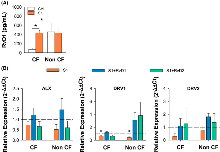
FIGURE 2.

SARS‐CoV‐2 stimulates RvD1 biosynthesis. RvD1 concentrations in MΦ cell supernatants following stimulation (3 hours) with CoV‐2 proteins (10 µg/mL) and in unstimulated cells used as a control (Ctrl). RvD1 was measured using a validated EIA procedure. 5 Results are mean ± SE from experiments with cells from four different donors. *P < .05 (One‐Way ANOVA). B, Real‐time PCR analysis of ALX, DRV1, and DRV2 receptors in CF and non‐CF MΦ treated (3 hours, 37°C) with S1 (10 μg/mL) plus RvD1 (10 nM), RvD2 (10 nM) or Veh (0.01 % EtOH). Results are mean ± SE of experiments from three different donors. Gene expression was determined as a duplicate for each test condition. *P < .05 (One‐Way ANOVA). ALX, lipoxin A4 receptor; DRV1, RvD1 receptor; DRV2, RvD2 receptor
