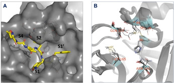
Figure 2.

(A) Surface representation (gray) of the substrate binding site of aligned Mpro used in ensemble docking, with designated subsites S1, S1’, S2 and S4. Ligand (gray sticks) and surface are obtained from PDB:6LU7. (B) Aligned Mpro structures used in ensemble docking. The residues involved in detected binding site flexibility are depicted in black lines and labeled with red letters.
