
Figure 1.
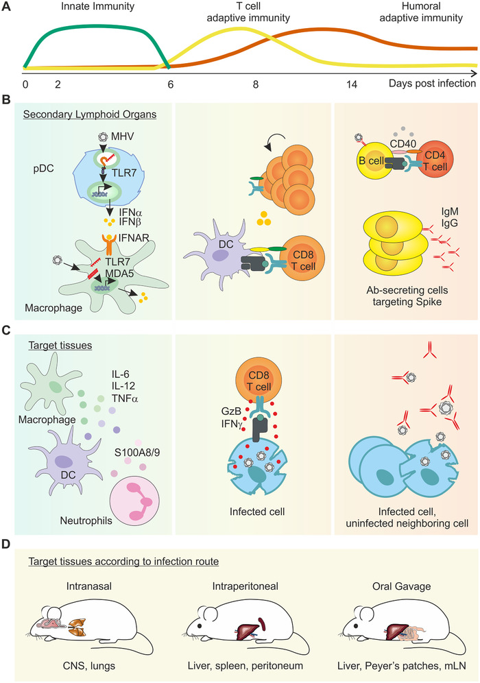
Schematic diagram of the overlapping layers of protective immunity against murine coronaviruses. (A) Schematic diagram of the timely overlap of innate and adaptive immune responses to MHV‐A59. (B) Virus‐infected cells rapidly produce type I IFN as the first line of innate immune defence. Plasmacytoid dendritic cells (pDC) residing in secondary lymphoid organs are a dominant source of type I IFNs, and signalling via the IFN‐α/β receptor (IFNAR) on macrophages triggers downstream innate mechanisms to limit viral spread. Uptake of viral antigen and activation by dendritic cells (DC) instigates cytotoxic CD8 T‐cell priming. Germinal centre B‐cell responses also take place in secondary lymphoid organs, promoting the expansion and maturation of high affinity, antibody‐secreting cells. (C) In the target tissue, cytokine‐secreting, activated myeloid cells help contain viral spread until primed, cytotoxic CD8 T cells migrate to the site of infection and critically eliminate virus‐infected cells. Long‐term humoral immunity is mediated by neutralizing antibodies (Ab). (D) Schematic diagram of the known target organs following MHV‐A59 infection according to distinct infection routes.