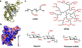
Figure 2.

a) Crystal structure of the SARS‐CoV‐2 spike protein RBD (PDB ID: 6M0J) [27] with a few important cationic residues that interact with polyanionic ligands. b) The electrostatic potential map of RBD. c) Schematic illustrations of polyglycerol sulfates in linear and hyperbranched architectures, and of the natural polysulfates, respectively. The negatively charged groups are marked red.
