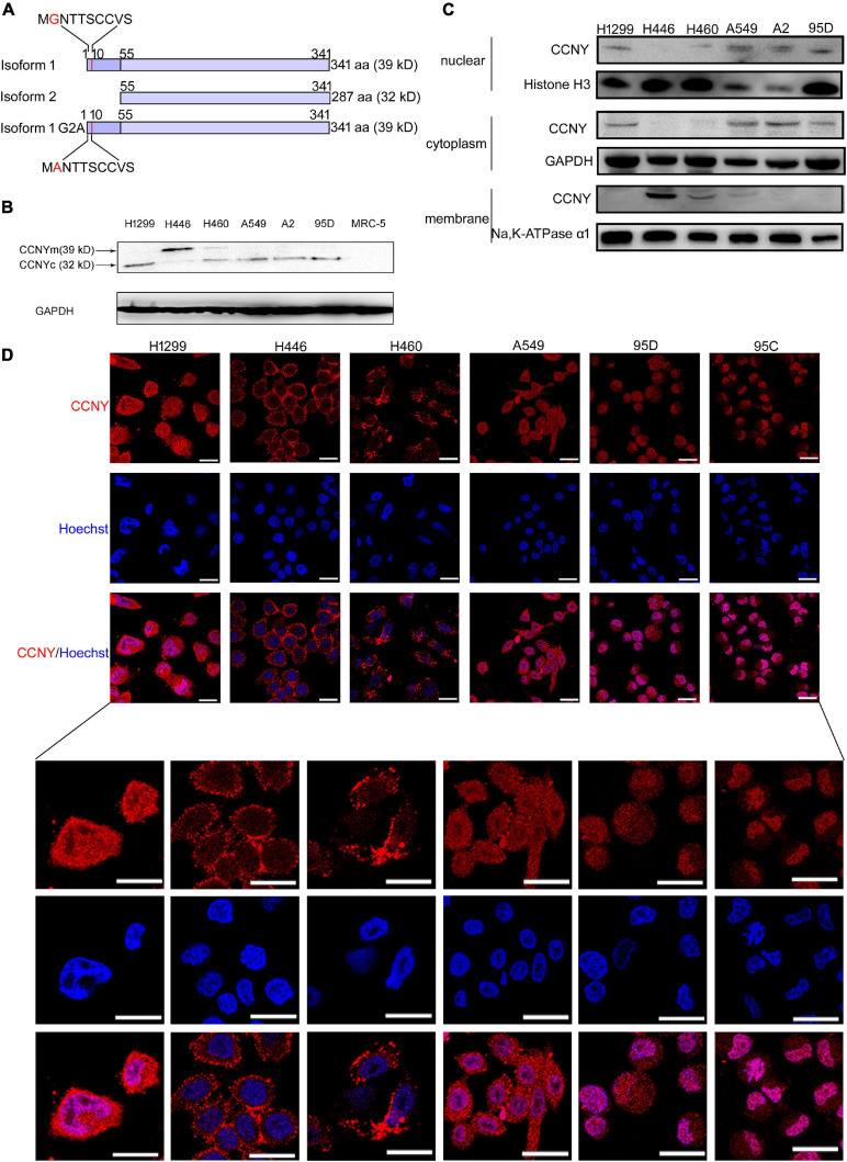
FIGURE 1.
CCNYm and CCNYc were highly expressed in lung cancer cells. (A) A simple view of the amino acid sequence for CCNY isoforms. The letter G with red color in the CCNY isoform one sequence is the myristoylation signal motif. (B) The levels of CCNY and loading control GDPAH in lung cancer cells and MRC cells were determined by WB. (C) Western blot was done using cell cytoplasmic extracts, membrane extracts and nuclear extracts, respectively. The CCNY level in cell membrane, cytoplasm and cell nucleus was shown in the figure. (D) Intracellular localization of CCNY in lung cancer. Bars represent 20 μm. The red fluorescence represents CCNY protein, and the blue fluorescence is the nuclear DNA staining with Hoechst 33342.

